You’re a what?… I’m a medical writer (oh, and I’m in NZ)

Imagine the scenario… you’re at a party and you’re introduced to someone new. Exchanging pleasantries, you’re asked THE question you hate to answer. You start to twitch, stutter, and eventually you say, ‘I’m a medical writer…’. Before you’ve finished saying the words, you already know their reply; in fact, you can – if you’re on the ball – mime the response at the same time as they say it (but that would be rude…) ‘YOU’RE A WHAT?’

That’s right, I’m a medical writer! I write about medical things… there’s more to it than that, but that’s how my explanation often starts. However, if I’m a little more alert, it seems the best way of explaining what I do, is to explain where I came from…

…Always a geek and at the age of 11, I knew I wanted to  be a cancer researcher. Why? My Grandad died of it, from this personal experience, I could see cancer REALLY sucks – I wanted to bosh it on the head. Simple. A few years’ of education ensued (and a few more)… I did a degree in Biology, a PhD in cancer research, and postdoctoral research in leukaemia. I loved it, but at the time I was 26 and saw too many people 10 years older than me on 1-month rolling contracts. The instability was not for me so I got out as soon as I could and moved into medical communications.

Medical commucations, or medcomms, as those in the know call it, is a thriving industry full of spods such as myself who support doctors, nurses, patients, and pharmaceutical companies in all of their medical writing activities. From writing a scientific manuscript for a journal such as The New England Journal of Medicine, through to writing a patient counselling tool, or even helping doctors with scientific presentations at big international meetings – a medical writer does allsorts! I love what I do. Someone else does the research, and I help them write it up! I’m a professional medical writer (see this paper for a down and dirty explanation of professional medical writing). I’ll write more about this another time. But this is how I ended up in New Zealand. It turns out medical writers are lacking over here, and New Zealand Immigration recognised that and gave me a residency visa (a.k.a my golden ticket) that allowed myself, my lovely wife, and The Spaniel to move New Zealand.

To give you an idea of what I do, here is a lightly edited version of a post from a recent initiative by a cool guy called Peter Llewellyn in the UK, which plotted A Day in The Life of Medcomms as it happened throughout the world. Hopefully it will provide you with a taster of what I do.

“Imagine… it’s a dank, dark, day, its mid-January 2011, and you’ve worked over Christmas as part of the run up to American Society of Clinical Oncology (ASCO) submissions (or FI-ASCO as I endearingly call it) [for readers not familiar with it, this is the largesst cancer conference in the world]. Your client is on the West Coast of the US and you’re working until silly o’clock again for the nth time in a row to cover the US time zone.

Well, that’s how my journey to Auckland, New Zealand began. I will spare you the full details of my journey (for now…), but working long nights was getting pretty tedious, and the solution seemed simple – flip time zones! I could then start work earlier in the day and finish earlier! I guess that’s the magic of MedComms, with a laptop and a phone you can work almost anywhere with a few minor exceptions (Antarctica anyone?).

At 7:30 am this morning, I checked my emails from home. My clients are a US MedComms agency, and so given the overlap in time zones, there is a window of opportunity up to 11am New Zealand time to read and respond to any emails, or chat to the client if needed. And then it was off to the beach to walk the dog! Auckland has fantastic beaches, and even in the middle of winter it is pretty mild. This also gives me the opportunity to mull over my work for the day, and consider what my priorities should be.

At 8:30 am(ish), suitably refreshed after walking the dog and a short trip to the office, I got straight into my day of medical writing. I received a brief overnight to write an outline for a manuscript for a top-tier general medical journal. First, I listened to a recording of the kick-off call with the authors and client, and the rest of my day was then spent writing the outline. The great thing about working in New Zealand is that I can do the part of a medical writing job I REALLY enjoy… the writing! For the client, having a writer in New Zealand allows projects to be kept moving forward around the clock. They can effectively work all 24 hours in the day, and no-one drops through exhaustion.

As your working day begins, I’m now on the beach (see below). I’ve now handed the baton over, and it’s now up to you to keep that MedComms wheel spinning, until tomorrow that is…”

Image

Possible Link Between Childbirth And Risk of Certain Types of Breast Cancer?

Giving birth may influence the risk of getting certain types of breast cancer according to recent research

When a breast cancer patient has their tumor biopsied or removed, a pathologist will take a sample of the removed cancer cells and will test them for receptors for estrogen, progesterone, or for HER2. The results of these tests will help determine the best treatment for the patient and the overall prognosis (or chance of recovery).

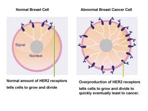
Receptors are proteins that help cells communicate with the outside world. When a receptor binds a specific molecule, various processes within the cell are triggered. For example, when estrogen binds to estrogen receptors and progesterone binds to progesterone receptors normal cellular processes involved in cell growth and division are triggered. However, in breast cancer, one of both of of these receptors  are overproduced triggering breast cells to grow and divide rapidly, which eventually leads to the development of a tumor or cancerous growth.  Relatively recently it has been discovered that another receptor, HER2- which seemingly does not have any molecules which bind it naturally- is also overproduced in some breast cancers.  HER2 overproduction also tells breast cancer cells to grow and divide rapidly, also leading to tumor growth. 

Most breast cancers have one or more of these receptors; however one type, called triple negative breast cancer does not have any of them.

Tumors that that have estrogen receptors generally respond well to treatment with antiestrogens, which prevent estrogen binding the estrogen receptors and triggering growth of breast cancer cells. Likewise, tumors that have HER2 generally respond well to trastuzumab, more commonly known as Herceptin, which stops HER2 triggering growth of breast cancer cells. However, triple negative breast cancers seem to be more difficult to treat and has a higher chance of the cancer coming back (recurrence). In addition, a large proportion of these tumors contain the BRCA1 mutation; a genetic fault which generally signifies an increased risk of a women getting breast cancer in her lifetime.

Hormone levels change during pregnancy, with estrogen being produced by the placenta throughout a pregnancy until birth. Because triple negative breast cancers, don’t have any estrogen receptors and hormone levels change during pregnancy, it is thought that childbirth could influence the risk of getting this type of breast cancer.

To address this question, Phipps and colleagues at the Fred Hutchinson Cancer Center in New York used data from over 150,000 women enrolled in the Women’s Health Initiative, a large study of postmenopausal women from 40 centers across the United States. 155,723 women were monitored over 8 years. Of these women, breast cancer developed in 6,194 women and complete data on estrogen receptor, progesterone receptor, and HER2 status was available in 307 women who developed triple-negative breast cancer, 2,610 women with estrogen receptor positive breast cancer, and the remaining cancers were estrogen receptor negative/progesterone receptor positive or estrogen receptor negative/progesterone receptor negative/HER2 positive (n=154). 150,529 women were used as controls.

They found that women without children had an increased risk of estrogen receptor positive breast cancer, but decreased risk of triple-negative breast cancer compared to those who had given birth to children. Specifically, having not given birth to children was associated with a 39% lower risk of triple negative breast cancer, but a 35% higher risk of estrogen receptor positive breast cancer compared with women who had given birth to children. For the statistically minded, see full statistics below. However, of those women who had given birth, those with more children had a higher risk of triple-negative and lower risk of estrogen receptor positive breast cancer (see below).

The authors note that they only studied postmenopausal women, so the findings reported here may not apply to younger premenopausal women. The findings should also be approached with considerable caution, as the study used a relatively small number of women so results need to be confirmed with a larger proportion of women. In essence, this is an interesting hypothesis-generating study (a study which scientists will use to further evaluate the influence of having children on the risk of having certain types of breast cancer), if indeed having children does influence the risk.


Reference: http://jnci.oxfordjournals.org/content/early/2011/02/23/jnci.djr030.full

For patient information: please see:

  1. http://www.cancerhelp.org.uk/about-cancer/cancer-questions/triple-negative-breast-cancer
  2. http://www.cancer.gov/cancertopics/pdq/treatment/breast/Patient/page1

Twinkle, Twinkle, Little Star…

If you don’t know already, Tefalhead is a medical writer, tinkering away at his keyboard writing ‘stuff’ for other folks. Most of his writings are based on clinical trial data for whatever product he is working on, and for whomever chooses to use his writing talents; usually a pharmaceutical company. As a result, Tefalhead does not have much artistic license when it comes to inventive manuscript titles. But, Tefalhead has a burgeoning artistic side, which is often desperate to show itself (in a non Jekyll and Hyde kind of way, it’s ok folks…).

Until now Tefalhead had gotten used to writing according to CONSORT guidelines, guidelines for transparent reporting of medical trials. But Tefalhead often muses on the lack of opportunity to write creative titles in his medical manuscripts. While thinking this, Tefalhead cast back his mind back to years gone by when he was a lonely, nerdy, little PhD Student, residing in Dallas, at the University of Texas Southwestern Medical Center. Here, the research was exciting, bold, inventive, creative even… and it is this creativeness that often ended up in manuscript titles, such as this….by Ron Estabrook and Bill Rainey:

Boy, these guys were good – gods in Tefalhead’s scientific world. In describing how one protein, called StAR controls how  hormones are produced in the cell, they don’t just stop with the imaginative title. Oh no, check out the last sentence…. “…and colleagues have demonstrated that StAR will continue to shine—but a twinkle remains in the eyes of those pursuing the detailed mechanism through which this long sought labile protein participates as an acute regulator of steroidogenesis.” So much better than the star trek sounding ‘New frontiers in…’ or the really uninviting ‘A perspective on…’, and leaves you with a pleasant picture of a twinkle in someone’s eye…. (mmm…)

Proof scientists are creative beings indeed… but that article is from 1996- eon’s ago in the scientific world! Have any other scientists demonstrated artistic flare in their manuscript titles? Well, it seems so… Tefalhead did a quick literature search and quickly found these little gems…

Through a glass darkly: advances in understanding breast cancer biology

Presumably in reference to the Bible passage from 1 Corinthians 13, in which seeing through a glass darkly refers to our understanding of God when we are alive, but the view will only be clear when we die. When reading this Tefalhead found his head slunking over his keyboard in a wave of despair… boy, this sounds depressing given we are talking about developments in breast cancer biology! Tefalhead reckons the authors presumably wanted to convey the fact that our vision of breast cancer at the moment is an imperfect or obscure vision of what is ‘reality’. Not so depressing I guess… hey, these guys are on another level! Here is another…

Transporters, Trojan horses and therapeutics: suitability of bile acid and peptide transporters for drug delivery

 Sounds like a bit of The Matrix mashed up with a bit of greek mythology doesn’t it?

We’ll ignore all reference to ‘Achilles Heel’… you guys are wasters, but Tefalhead reckons there are lots of opportunities for you scientists out there to be creative in writing your manuscript titles, and is a little jealous, after all it’s little restrictive when all you really have to play with is…

Phase 3 randomized controlled study of [insert drug] in patients with [insert disease]…  yada yada yada

But, hey, who said being a medical writer was all rock and roll! (But I like it…)

Chronic Fatigue Syndrome: Stop PACEing Around and Move On?

Tefalhead is not an angry man, in fact, Tefalhead is a mild-mannered medical writer who nonchantly keeps on plodding on in his own Tefalhead world; indifferent to most happenings in the world. But, that all changed last week when he saw headlines like the one below:

Tefalhead was not the only one to be stirred by the release of the PACE(Pacing, Activity, and Cognitive behavioural therapy, a randomised Evaluation) study of various treatments for myalgic encephalomyelitis (M.E)/chronic fatigue syndrome (CFS) in the Lancet this week. The Twittersphere, and the internet as a whole was full of chatter spinning off in every direction following on from the PACE study publication, with blog titles such as “Invest in ME: PACE trials are .Yup, that’s right claims that a study published in the prestigious journal The Lancet’ are BOGUS! Controversy surrounding the study was not confined to random bloggers, tweeters, or such like but the M.E. Association had this cracking press release:


 Yup, you read it correctly “Results are at serious variance to patient evidence…“. Let me get this right, one of the main M.E/CFS patient advocacy groups in the UK disagree with the results published in the Lancet, one of the “world’s best known, oldest, and most respected general medical journals“ according to Wikipedia!

 It’s now Sunday, and Tefalhead has recovered from the red mist that blinded his thoughts on Friday. Tefalhead is a scientist by training and although he desperately wanted to blog about this on Friday, he knew his scrawlings would lack objectivity. However, after two days of chillin’, his objectivity head is back on, but first, Tefalhead has one thing to disclose…nope, we’re not talking about receipt of consultancy fees from The Department of Health, or that Tefalhead is one of the co-authors’ of the study. No, this is much more personal. Tefalhead’s wife has M.E/CFS a condition that is misunderstood by all, massively impacts lives of those afflicted with this condition and has no known magic bullet in terms of a cure. In brief, this condition sucks BIG time- and to see the love of your life affected by it – well, there are no words for it. Needless to say, because of this fact, there will be inherent biases in what I repor. It’s inevitable folks- so treat what I say next, with CAUTION.

What did the study involve?

The study was carried out by researchers from several UK institutions, and was funded by the UK medical research council, The Department of Health for England, the Scottish Chief Scientist Office, and The Department of Work and Pensions. The study was published in The Lancet online this week at http://www.thelancet.com/journals/lancet/article/PIIS0140-6736(11)60096-2/fulltext . Whoa, right, let’s backtrack for one second…. “funded by… The Department of Work and Pensions’? For non-UK readers, the Department of Work and Pensions is an UK government department that manages welfare and pension policy in the UK, including provision of financial support for people claiming disability benefits. So what on earth are these guys doing funding a clinical study to assess the effectiveness and safety of treatments for patients with M.E/CFS.? Hmmm… Tefalhead, asked the very same question. After digging around a bit, he stumbled across this document http://www.meactionuk.org.uk/magical-medicine.pdf from Malcolm Hooper, an Emeritus Professor Of Medicinal Chemistry…. This document seems to open up a whole lot of controversy surrounding the PACE study, including suggestions that the Department of Work and Pensions could use the data from the study to essentially remove many CFS patients from benefits system in the UK. That would save some money right? Not a bad thing in this era of austerity measures. Tefalhead does not want to distract from the main analyses reported in the Lancet study, but the fact that an external body that has inherent interests in the data reported actually funded the study… is a little bit fishy. However, it happens all the time with pharmaceutical companies for example, who are continually reporting the results from their clinical studies in manuscript articles. When reading such articles one needs to take into account the phenomenon of funding bias and acknowledge that the conclusions of a study could be biased towards the outcome the funding agency wants. To be clear, Tefalhead is not saying the funding bias has occurred in the PACE Lancet study, but we need to take this into account when considering the data.

So… where did this study come from?

Details of the Study Design are given in Box 1. In brief, 641 patients were given one of four treatments: specialist medical care (SMC)(provided by doctor with specialist expertise in CFS), SMC plus adapative pacing therapy (aims to assist CFS patients in optimally using their limited stores of energy); SMC plus cognitive behaviour theapy (CBT; believes that fear responses are linked to physiological response and make the fatigue worse and involves strategies such as establishing a healthy sleep patterm addressing fears, problem solves and working with a therapist to increase mental and physical activity), and SMC plus graded exercise therapy (GET; believes that fatigue can be reversed or reduced using gradual increases in activity and uses strategies to establish a baseline of acheivable exercise followed by negotiated incremental increases in the time spent being physically active).

Patients were given questionnaires to fill in to assess the effectiveness of treatment and were unmasked to treatment, ie it was obvious what treatment they were receiving. As a result, their own expectations of what treatment they were receiving, could have influenced the final results. To account for this, the investigators did assess patient  expectations before treatment started. The study clinicians also used questionnaires and scoring methods to assess the impact of treatment on the patients’ overall health, Any adverse events were also monitored forthroughout the study (any adverse change in health or side effect during treatment).

There were no biological measures assessed during the study, which would have been expected for a condition such as CFS where the biological basis of the condition is unknown. Tefalhead thinks this is a big shame given the large amount of patients involved in the study.

What did the researchers find?

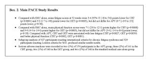
After 1 year, the study found that fatigue was significantly lower for CBT and GET compared with SMC alone (See Box 2). However, there was no significant difference between APT and SMC. Physical functioning (the ability to perform a range of activities from self care to more vigorous acitivities) was significantly higher for CBT and GET versus SMC, but there was no significant difference between APT and SMC. Compared with APT, CBT and GET were associated with less fatigue and better physical functioning. The Oxford criteria for diagnosi of ME/CFS are broader than other criteria used, and as a result, may cover a broader range of conditions that may be unexplained and thus categorized as ME/CFS. When different diagnostic criteria for CFS were applied (the international CFS criteria and the London criteria for ME), two-thirds of patients met the international CFS criteria and about half met the M.E criteria, which requite the presence of post-exertional relapse and excludes patients with depressive of anxiety disorders. When these new criteria were applied, the authors found similar results with no significant differences in results results between the different criteria used.

Of note, after treatment 16% of patients in the APT group, 30% of patients in the CBT group, 28% of patients in the GET group, and 15% undergoing SMC were within ‘normal’ ranges for the fatigue and physical functioning outcomes measured. Let’s switch that around, 70% of patients in the CBT group and 72% of patients in the GET group had abnormal levels of fatigue and physical functioning. In addition, a clinically useful difference in results for the primary outcomes equated to 2 points on the Chalder fatigue questionnaire and 8 points for the short form 36 questionnaire, ie it just took a small change for the therapies to be clinically useful. Using these relatively loose criteria, only 42% of patients on APT, 59% of patients on CBT, 61% of patients on GET, and 45% of patients receiving standard medical care ‘improved’  at 52 weeks.
 

How were the results interpreted by the authors?

The researchers conclude that CBT and GET can be safely added to SMC to moderately improve outcomes for ME/CFS, and that APT is not an effective treatment. They also state that findings were similar for patients meeting the different diagnostic criteria for ME/CFS , and for those with depression. Importantly, they note that there were no important differences in safety outcomes between treatment groups

Author conclusions aside, how relevant are these results?

 This study had many strengths including: (1) a low dropout rate in the study, which is impressive given that patients could drop out part way if they felt they no longer wanted to continue treatment; (2) there were high rates of patient satisfaction; (3) well defined treatment protocols were used to name but a few. However, there are several limitations of this study to consider:

  • Patients who were unable to attend hospital were excluded, so the results from this study cannot really be applied to ME/CFS patients who are bed bound (there are quite a few out there)
  • Standard medical care is not the same as the usual medical care given by a family doctor or GP
  • Children who are also afflicted by ME/CFS were not included in the study
  • Conventional criteria were used to define clinically useful differences between treatments
  • Masking of patients or clinicians to the treatment given was not possible given the types of treatment given and research assessors were not masked, which may have led to reporting bias

One major objection to the study seems to be that the criteria used to select ME/CFS patients does not accurately reflect the ME patient (again see the response from Malcolm Hooper here). However, the authors do apply the results to alternative criteria for diagnosing CFS. Although this is no substitute for using the other ME/CFS criteria for inclusion into the study.

Perspective from Action for ME

Action for  ME, a UK-based patient advocacy group have serious concerns about the PACE study, stating “The largest ever clinical trial into the effects of CBT, GET and adaptive pacing therapy (APT) has produced results that are clearly at serious variance from those reported by the largest ever survey of patient opinion on these forms of treatment.

We find the trial results extremely worrying because pacing, in the form that the MEA recommends, may as a result no longer be offered as a treatment option in NHS clinics. And at the same time, NICE may well strengthen its inflexible and unhelpful recommendations regarding CBT and GET.

We also fear that the way in which the results are already being reported in media headlines – eg Got ME? Just get out and exercise, say scientists – will lead some doctors to advise inappropriate exercise regimes that will cause a serious relapse.

This is not a good day for people with ME/CFS.

They have a complex multisystem illness that requires a range of treatment options based on their individual symptoms as well as the stage and severity of their illness”

So, there you go, Action for ME have a lot of serious concerns not only about the design of the study, but also the implications for ME/CFS patients throughout the UK. 

Stop PACEing Around and Move On

The results from this study highlight the plight of patients with ME/CFS, and should fuel further research into the basic biology of what causes ME/CFS, together with clinical studies to determine what treatments really are effective for these patients. The lack of any harms with any of these treatments, should allay patient fears, that some of the treatments can do them harm. However, any confusion in findings can only be bolstered by rigorously designed clinical studies.

Tefalhead just hopes that something can be done to help his wife regain some type of  ‘normality’ in her life, and is reminded that it isn’t just numbers reported in this study, but results from patients whose lives are hugely affected.

There is a great deal of disunity amongst ME/CFS patients, clinicians and patient groups, and it seems that the main people losing out here are the ME/CFS patients themselves. Tefalhead is at a loss as what to suggest in terms of a way forward… thoughts anyone? However, a line clearly needs to be drawn under the furore of the PACE study, and a fresh start made [Tefalhead suggests a group hug, but that may not be everybody’s idea of a good time].

Tefalhead has one last little grumble…headlines such as ‘Got ME? Just get out and exercise”. Are not only irresponsible but downright dangerous. Tefalhead spoke to a leading expert in the ME/CFS field at the weekend, who reiterated that any exercise therapy must be done under medical supervision. Scientific journalists- careful reporting PLEASE. If your headlines are taken literally by vulnerable patients, serious harm could be done.

Note from TefalHead: Your comments on this topic are appreciated, BUT please remember Tefalhead is as concerned with this issue as you are, and he is just trying to put the topic out there for debate. In other words… be nice, thanks. Oh, and Tefalhead has purposefully steered well clear from the XRMV issue, mainly to report the results of the PACE study and not distract from that.  Tefalhead will review the XRMV story at some point in the future


Human Enzyme Made in Plants May Protect Against Nerve Agents

On Monday March 20, 1995, 5 members of the Japanese cult, Aum Shinrikyo walked onto their appointed trains with deadly parcels of the nerve agent Sarin wrapped in plastic bags and newspapers. At prearranged stations, the sarin packets were dropped and stabbed with umbrella tips to release deadly sarin vapours. Twelve people were killed, and nearly 5,000 were hospitalized.

As a result of the rise in global terrorism such chilling attacks remain a distinct possibility. However, the means to fully treat patients remains elusive as current anti-nerve agent treatments can only help symptoms but do not prevent long term nerve damage.

The human nervous system is constantly chattering away with the firing of impulses along lines of interconnected nerve cells. Signals are sent from the brain to various parts of the body through firing of impulses from one nerve cell to the next, and also from nerve to muscle cells which then results in a muscle contraction, for example contraction of muscles associated with breathing. Transmission of impulses from nerve to muscle cells requires the release of a substance called acetycholine at the end of the nerve cell which is immediately broken down by acetycholinesterase (AChE) into inactive components. The immediate breakdown of AChE is essential for coordinated movement of muscles. If this does not occur, acetycholine floods the nervous system resulting in paralysis and disorganized muscle contractions eventually leading to death when muscles required for breathing and heart muscles are affected. This occurs with nerve agents which prevent the breakdown of AChE.

Butyrylcholinesterase (BChE) occurs in blood, scavenging and binding to toxic nerve agents, such as sarin gas to prevent them from doing their dirty work of stopping the breakdown of AChE. However, there is not enough of BChE within the blood for it to be mass produced for stockpiling for the next bioterrorism attack, and of course there is the risk of transmitting blood disease- think back to the haemophilia scare of the 1980s when contamined blood products resulted in patients being needlessly infected with HIV.

However, recent research by Brian Geyer and colleagues in the November issue of The Proceedings of the National Academy of Sciences suggests an alternative for mass production of BChE to treat nerve agent attacks. They used tobacco plants which were genetically modified, also known as transgenic plants, to over produce BChE in their leaves. In their experiments Geyer et al. showed that this plant-derived BChE protected lab animals exposed to nerve agents and alleviated symptoms of poisoning such as difficulties in breathing and convulsions, and also prevented death. They also found that they could make the enzyme persist in the blood of the lab animals for longer by binding it to the chemical polyethylene glycol (PEG) to allow for further protection.

Much work remains before plant-derived BChE can be used to protect humans against nerve agents, but the fact that it can be produced in plants and demonstrates promising signs of benefit in lab animals is tantalizing.

As transgenic plants could be an abundant and relatively inexpensive source of BChE, they could be used for large-scale production and stockpiling of this treatment for the likely event of another terrorism attack.

Reference: http://www.pnas.org/content/early/2010/11/05/1009021107

Don’t Stress – Just ASK!: New Role Found for Stress-Activated Molecule in Preventing Cellular Dustbins from Degrading Waste

The stress-activated molecule, ASK, prevents cellular dustbins from degrading waste proteins and may offer a new target for treating various diseases including cancer, AIDs and neurodegenerative diseases, according to a study in the Journal of Biological Chemistry this month.

It happens to us all, you’ve got a ridiculously tight deadline, an exam tomorrow, or something else that really stresses you out, and your heart starts beating a little bit faster, your palms get sweaty, and you feel sick. These responses are some of the direct results of stress on the human body. Similarly, just as the body as a whole can react to stress, so can individual cells.

ASK1 is triggered when a cell is exposed to stressful stimuli including oxidative stress (when toxic free radicals are produced within the cell which can cause serious damage to DNA) or endoplasmic reticulum stress (accumulation of unwanted proteins within the endoplasmic reticulum [the area of the cell responsible for protein production]). When triggered, ASK1 translates the stimuli of stress to within the cell, activating various intracellular processes linked to cell death and normal cell function. However, recent research by Um and Im in the November issue of the Journal of Biological Chemistry have found an additional role for ASK1 in that it inhibits the actions of cellular dustbins, or ‘proteasomes’, which eat up unwanted proteins in cells, a process which is vital for normal cell function.

Proteasomes break down unwanted proteins by a process called proteolysis to smaller components called amino acids, which are then re-used to make more new proteins. These unwanted proteins are tagged by a small protein called ubiquitin. Once tagged, these proteins are doomed as the cell is alerted to add additional ubiquitin molecules, and degrade the tagged protein. Well, that’s the norm, but as with most diseases sometimes something goes wrong and proteasomes  can malfunction. As a result, some proteins essential for normal cell function, are lost or accumulated depending on whether protein degradation is increased or decreased. This can lead to diseases such as cancer, AIDS, and neurodegenerative diseases .

In a series of elaborate and very thorough experiments, Um and Im investigated the link between ASK1 and proteasome function (these guys were meticulous; in fact Tefalhead reckons the whole set of experiments took 2 to 3 years!).

They specifically chose to work on the 26S proteasome, the most common proteasome in human cells, which is composed of two 19S ‘lids’ where the proteins are tagged for destruction and the 20S hollow core where proteins are mashed up (Figure 1). Hey, like I said earlier, this thing really is a cellular dustbin!

Figure 1. Parts of the 26S proteasome- a cellular dustin

They found that ASK1 interacts with the 19S lids of the 26S proteasome, the parts responsible for tagging unwanted proteins and transferring them to the core to be mashed up. Specifically ASK1 seems to inhibit an enzyme in the 19S lid called ATPase Rpt5, which is  critical for normal 26 S proteasome activity.

The authors go on to hypothesise that ASK1’s ability to inhibit 26S proteasome activity through blocking ATPase Rpt5 may account for the reduction of 26S proteasome activity in stressful conditions in the cell, which can eventually lead to disease. If scientists can develop inhibitors of ASK1 to stabilize proteasome activity, and if any beneficial effects are translated from the bench into the clinic, they may provide much needed treatments for cancer, AIDs, and neurodegenerative diseases.

Lots of ‘ifs’, but only time will tell whether ASK1 is a new target for the future

Ref: http://www.jbc.org/content/285/47/36434.abstract

‘Silent’ Cytomegalovirus Can Cause Intestinal Cancers in Mice: Significance in Humans Still Unclear

Figure 1. The CMV virus, which occurs in up to 90% of the worlds' population

Human Cytomelagovirus (CMV), which infects millions of people worldwide usually without any symptoms, appears to cause gastrointestinal cancers in mice, the significance in humans is not clear.

Up to 90% of the worldwide population are silently infected with CMV (Fig. 1), which generally is not associated with any symptoms, but is a persistent little blighter nonetheless. CMV infection can cause severe disease particularly in people with a weakened immune system such as transplant patients, and AIDS patients, and  developing fetuses. People with a healthy immune system can mount a vigorous immune response to keep the virus at bay; however, in people with a weakened immune system, the virus can rage out of control and cause severe disease which can be life threatening.

 Unlike other organisms, viruses do not replicate through cell division; instead they invade and exploit host cells to do their replication for them. To do this, they must first bind to a host cell, enter it, and then hijack the hosts’ cells machinery to replicate and produce more viruses in the so called ‘primary infection’ stage. CMV viral replication is typical amongst viruses in that during the hijack of a host cell, viral DNA integrates into host cell DNA resulting in the production of various genes needed for viral replication. There are several phases of gene expression: immediate-early, delayed early and late, based on the time from initial viral infection. Following primary infection, CMV can also enter into a latent phase when the virus enters a dormant state during which viral replication does not occur. However, CMV can be later be reactivated back to a replicative state which can occur in people with a weakened immune system.

During the immediate-early gene and latent phases  of a CMV infection a gene called US28 is produced. As CMV infection had previously been detected in the cells that line the gastrointestinal tract and since the role of US28 has only been investigated in in vitro studies, Bongers et al., chose to examine the role of US28 in tumor formation in cells lining the gastrointestinal tract of mice, as reported in this months’ issue of the Journal of Clinical Investigation. To do this, they created transgenic mice─ mice where extra genes are added to the genome (Fig. 2)─ in this case the US28 gene was specifically targeted to cells in the lining layer of the mouse gut [Fig. 3]) and the lining layer of the gut was examined to see if there were any cancerous changes.

Figure 2. Creation of US28 transgenic mice

Figure 3. US28 expression in the gastrointestinal lining layer of US28 transgenic mice

In US28 transgenic mice, they found that US28 had the ability to induce cancer by increasing cell proliferation in the cells lining the gut through activating of specific intracellular signaling molecules (wnt and MAPK). US28 transgenic mice also went on to develop intestinal cancers.

As patients with inflammatory bowel disease have an increased risk of developing colon cancer, they examined whether CCL2, which is expressed in many inflammatory conditions and inflammatory bowel disease, increased the ability of US28 to induce cancer which it did.

The authors conclude that US28 produced by CMV promotes the development of cancer in transgenic mice and suggest that CMV infection may facilitate the development of gastrointestinal cancers in humans. In addition, inflammatory factors such as CCL2 can increase the oncogenic activity of US28. Although this is the first in vivo study of it’s type and builds on previous in vitro studies which have implicated US28 and CMV in the development of cancer, it falls way short of drawing any firm conclusions that CMV could cause cancer in humans. In fact, drawing any such conclusions would be unwise as this is more of a hypothesis-generating study which could form the basis of further research to analyse the association between CMV and intestinal cancer in humans.

Reference: http://www.jci.org/articles/view/42563/pdf

Young and Old Mice Differ in Energy Regulation: Implications for Childhood Obesity?

An obese mouse unable to produce leptin compared to a normal mouse

The fat-busting molecule, leptin, appears to work in different regions of the brain in young compared with old mice.

In the mid 90’s, the way scientists think about what causes obesity was revolutionized with the discovery of leptin, an appetite-suppressing hormone.

Leptin is made by fat tissue and binds receptors in the brain to reduce energy intake by suppressing appetite and increasing activity to burn off energy, essentially regulating energy balance, or ‘homeostasis’.

Understanding how energy homeostasis is regulated is an area of intense research because of the current obesity epidemic. Importantly, childhood obesity is rising worldwide, together with associated conditions such as type 2 diabetes and heart disease. However, ways of treating childhood obesity remain mainly ineffective.

Leptin contributes to the regulation of energy homeostasis by acting on neurons in different brain regions, but exactly what effects each region mediates has not been clearly determined.

To address this question, Ring and Zeltser from Columbia University in New York, created mice in which leptin signaling was disrupted in only the hypothalamus of the brain (LeprNkx2.1 KO mice). When they compared the characteristics of these mice at a young age (<8 weeks) to mice lacking leptin signaling in all body cells (Leprdb/db mice), they found they were similar showing increased weight gain and increased amount of fat tissue. In contrast, after 8 weeks they found that mice with disrupted leptin signaling in the hypothalamus maintained consistent levels of fat tissue, whereas the mice lacking leptin in all body cells, became more and more obese. These findings suggest that leptin signaling in the mouse hypothalamus is needed to stop the development of fat tissue in young mice, whereas leptin signaling limits the development of fat tissue in older mice occurs through other regions of the brain.

The authors highlight that ‘these observations are consistent with the idea that the regulation of phenotypes related to energy homeostasis may be different (and less complex) in immature animals.’

The authors conclude ‘If this notion proves true, manipulations of critical components of circuits that establish metabolic profiles in young animals would represent a promising strategy to combat childhood obesity.’

Reference: http://www.jci.org/articles/view/41985

Bone Drugs May Thwart Development of Cancer ‘Seeds’

What do cancer cells and seeds have in common? The way that they spread to a new site and germinate it seems.

When a plant goes to seed, its seeds are carried in lots of different directions, but they can only grow and develop if they land on soil which is conducive to growth. Similarly, it is now well known that when cancer spreads secondary tumors (or metastases) do not develop by chance, as some organs provide a more fertile soil for tumor growth than others. These ideas form a long-running hypothesis in cancer research called the ‘seed and soil’ hypothesis.

A recent editorial in the Journal of Clinical Oncology this week by Dr Michael Gnat, further reinforces this hypothesis for cancer development, and that a certain group of bone cancer drugs, bisphosphonates, might create an “unfavorable soil” in which the development of cancer cells is thwarted.

Bisphosphonates are the treatment of choice for preventing bone loss and fractures in postmenopausal women with osteoporosis. They are also being investigated as treatment for preventing cancer-induced bone loss in women with early-stage breast cancer. Importantly, they have demonstrated anti-cancer activity in the lab and in clinical studies, which have shown that they:  block the release of factors that promote tumor growth and angiogenesis (growth of the tumors blood supply), induce cancer cell death (or apoptosis), prevent cancer cells sticking to each other, reduce the ability of cancer cells to spread via blood vessels, and activate the immune system to attack cancer cells. 

Based on this background information, several studies have been set up to determine whether bisphosphonates given to women with postmenopausal osteoporosis may reduce the risk of breast cancer, including two studies reported in the same issue of the Journal of Clinical Oncology.

In the first study by Chlebowski et al., approximately 150,000 women who received bisphosphonates for osteoporosis had a 32% reduction in the risk of breast cancer versus those who did not receive bisphosphonates. In a separate study by Rennert et al., a 28% reduction in the risk of breast cancer was reported in women receiving bisphosphonates for more than 1 year.

‘At this point, it would be premature to recommend the use of oral bisphosphonates to prevent breast cancer in all postmenopausal women. However, it is not unreasonable to consider the potential anticancer benefits of bisphosphonate therapy’

However, these results should be considered with caution as the authors note that several factors may have influenced the results, including age, ethnicity, and tobacco use to name but a few, says Dr Gnat. As a result, he states, ‘these analyses should be viewed as hypothesis generating and not practice changing at this time’…. ‘At this point, it would be premature to recommend the use of oral bisphosphonates to prevent breast cancer in all postmenopausal women. However, it is not unreasonable to consider the potential anticancer benefits of bisphosphonate therapy, in addition to its bone protecting effects’.

‘Bisphosphonate-induced changes to the microenvironment surrounding potential cancer cells can be exploited in preventing cancer’

Dr Gnat also states ‘The statistically significant reductions in breast cancer risk associated with bisphosphonate use “are profound and intriguing, because they suggest that bisphosphonate-induced changes to the microenvironment surrounding potential cancer cells can be exploited in preventing breast cancer,”. He goes on to say that these significant results are ‘profound and intriguing’ as they ‘suggest that bisphosphonate-induced changes to the microenvironment surrounding potential cancer cells can be exploited in preventing cancer’. He also suggests that these results support the idea that the seed and soil hypothesis is relevant to both healthy postmenopausal women as well as in preventing recurrence in women with early-stage breast cancer.

In conclusion, Dr Gnat suggests that future anticancer treatments may target the tumor microenvironment in addition to the cancer cells themselves.

References:

1. http://jco.ascopubs.org/cgi/content/full/28/22/3548

2. http://jco.ascopubs.org/cgi/content/abstract/28/22/3577

3. http://jco.ascopubs.org/cgi/content/abstract/28/22/3582

All Roads Lead to Blocking Androgen Signaling in Castrate-Resistant Prostate Cancer, But Which Road is Best?

Things are hotting up in the prostate cancer field, with scientists discovering several new targets that may provide hope for patients with late-stage prostate cancer.

Androgens (testosterone and dihydrotestosterone) are known to fuel the growth of prostate cancers as they bind the androgen receptor (AR) driving signaling pathways that driving tumor cell proliferation and tumor development. To halt the effects of androgens binding the AR, androgen deprivation therapy (ADT) is used to try and block the levels of circulating androgens and prevent them from fuelling tumor growth.

Despite ADT reducing androgen levels to very low or ‘castrate’ levels, castrate-resistant prostate cancer (CRPC) typically develops in most patients who initially respond to treatment. The precise mechanisms underlying this resistance are just starting to be worked out, but it seems that that ADT does not fully ablate androgen synthesis in the adrenal glands (where androgen synthesis normally occurs)  nor in the tumor itself where recently it has been discovered that androgen synthesis also occurs.

Once CRPC develops, the time to death is about 2 years and treatment options in this patient population are limited, demonstrating a high unmet need for treatments that prolong survival in these patients.

Androgens are synthesized from cholesterol involving complex enzymatic pathways, which includes the enzyme 3 beta-hydroxysteroid dehydrogenase which takes inactive precursors (DHEA and A5diol) and converts them to the more active androgens, androstenedione and testosterone, which are later converted to dihydrotestosterone. If this enzyme is active in prostate tumors, testosterone and dihydrotestosterone could in theory bind the AR and continue to fuel tumor growth. In a recent study in the journal Endocrinology, Evaul et al., discovered that was the case with 3 beta-hydroxysteroid dehydrogenase being necessary for producing testosterone and dihydrotestosterone in models of CRPC, which then activates the AR, driving tumor cell proliferation. As a result, this enzyme is thought to provide an attractive new target for the treatment of CRPC.

At the moment this is just a hypothesis that needs to be tested in the clinic. However, there are similar agents currently in later stages of development, including abiraterone and TAK700 which inhibit another enzyme in the androgen synthesis pathway (CYP17,20), MDV3100 which blocks androgens binding the AR, and VN/124-1 which both inhibits CYP17,20 and blocks androgens binding the AR. These are indeed exciting times for prostate cancer patients.

Reference: http://endo.endojournals.org/cgi/content/abstract/151/8/3514